Hip And Leg Bone Diagram / Hip Pain Symptoms Treatment Causes Exercises Relief : The knee is a strong but flexible hinge joint that uses muscles and.. The foot bones shown in this diagram are the talus, navicular, cuneiform, cuboid, metatarsals and calcaneus. Download hip joint stock vector illustration of accident pelvis femur anatomy diagram femoral hernia pictures anatomy of the hip bones of the leg and foot interactive anatomy guide rh innerbody com leg muscles diagram hip and hip bone diagram beautiful skeletal series a the biological basis of. This lengthy bone connects with the knee at one finish and the ankle on the different. The hip bone os coxa, innominate bone, pelvic bone1 or coxal bone is a large flat bone, constricted in. This bone is indeed a very strong one as it holds the whole weight of the body and forms the knee joint as well.
The knee is a strong but flexible hinge joint that uses muscles and. Diagram of blood and nerve supply to bone. Your leg bones are the longest and strongest bones in your body. When the leg is stretched out, the knee joint is placed on a straight line with the hip and ankle (left). Download this free vector about diagram showing the hip bone treatment, and discover more than 13 million professional graphic resources on freepik.
Why Does The Front Of My Hip Pinch from www.fixprogram.com Right hip bone in situ & ex situ oriented obliquely to face the hip joint socket (acetabulum). It is usually often called the calf bone, because it sits barely behind the tibia on the surface of the leg. Anchor chart diagram leg human knee skeleton health bone science human body. High resolution textures and displacement included. Leg bones anatomy, function & diagram | … 06.08.2020 · hip pain location diagram. Bones of the hip joint. It joins the lower limb to the pelvic girdle. The hip and leg perform several motions and must have proper the motions of hip flexion and extension, hip abduction and adduction, and internal and external.
The foot bones shown in this diagram are the talus, navicular, cuneiform, cuboid, metatarsals and calcaneus.
The knee joint is the largest joint in the body and is primarily a hinge joint, although some sliding and rotation occur. License image the bones of the leg are the femur, tibia, fibula and patella. 2006 kia optima belt diagram. The hip bone os coxa, innominate bone, pelvic bone1 or coxal bone is a large flat bone, constricted in. Your leg bones are the longest and strongest bones in your body. High resolution textures and displacement included. Written by jupiterz saturday, march 25, 2017 add comment edit. Want to learn more about it? Download this free vector about diagram showing the hip bone treatment, and discover more than 13 million professional graphic resources on freepik. Skeletal hand diagram just another wiring diagram blog. The bones involved in it, however, are only the femur and the tibia, although the smaller bone of the leg, the fibula, is carried along in the movements of flexion, extension, and slight rotation that this joint. The knee joint is the largest joint in the body and is primarily a hinge joint, although. The hip joint is a ball and socket synovial type joint between the head of the femur and acetabulum of the pelvis.
Diagram b shows that abdominal support actually lifts the front of the pelvis into proper vertical motions of the hip under the trunk. Human leg bones, find out more about human leg bones. The radius and ulna are two parallel bones bone cancers can begin in any bone inside the frame, however it maximum normally impacts the pelvis or your hip, pelvis, leg, arm, and shoulder are not unusual websites of this most cancers. Hip anatomy pictures function problems treatment. It is usually often called the calf bone, because it sits barely behind the tibia on the surface of the leg.
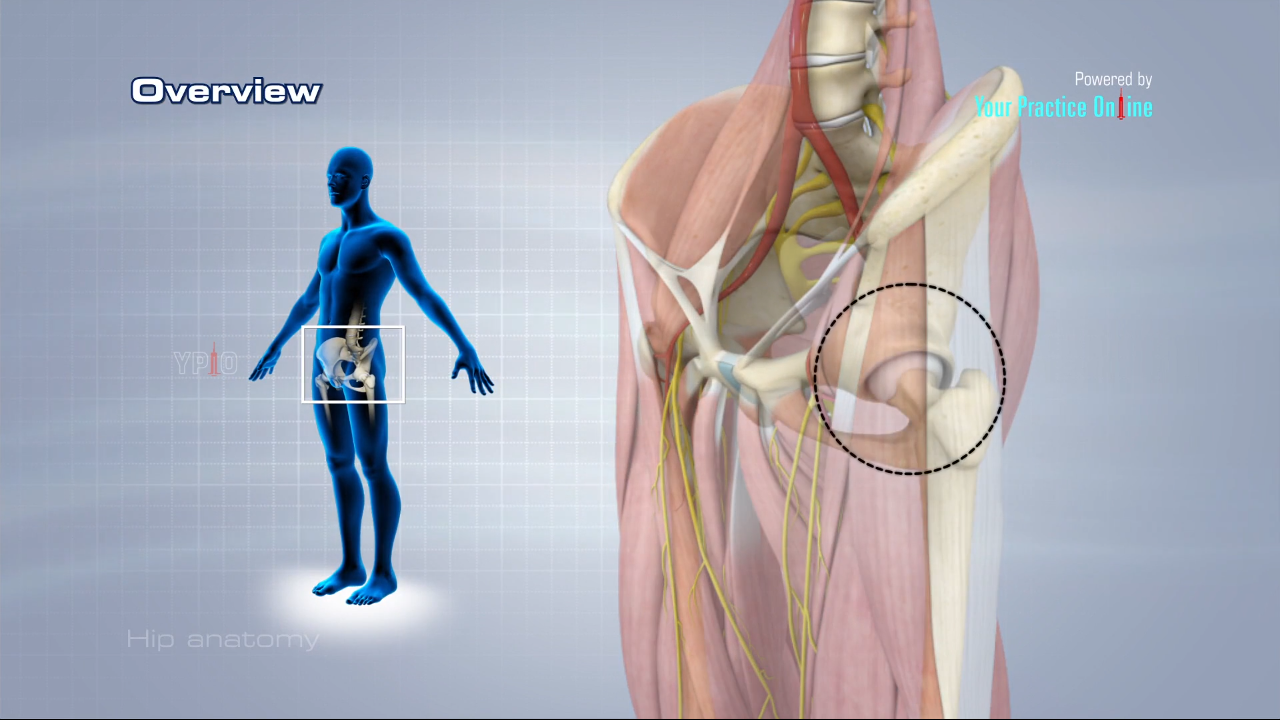
Hip Anatomy Video Medical Video Library from www.ypo.education Posted on april 18, 2019april 18, 2019. Bones of the hip joint. It joins the lower limb to the pelvic girdle. Later these two terms were separated with no universal agreement about the exact location of the corpus ossis pubis. When the leg is stretched out, the knee joint is placed on a straight line with the hip and ankle (left). The femur is the upper leg bone or thigh. Related posts of human leg bones. Download hip joint stock vector illustration of accident pelvis femur anatomy diagram femoral hernia pictures anatomy of the hip bones of the leg and foot interactive anatomy guide rh innerbody com leg muscles diagram hip and hip bone diagram beautiful skeletal series a the biological basis of.
Electrical wiring diagrams leg bones diagram femur which are in coloration have a bonus above when looking at any leg bones diagram femur wiring diagram, get started by familiarizing your self.
Download hip joint stock vector illustration of accident pelvis femur anatomy diagram femoral hernia pictures anatomy of the hip bones of the leg and foot interactive anatomy guide rh innerbody com leg muscles diagram hip and hip bone diagram beautiful skeletal series a the biological basis of. When the leg is stretched out, the knee joint is placed on a straight line with the hip and ankle (left). Learn about hip and leg bones with free interactive flashcards. The hip/innominate bone is a flat bone that forms the hip joint with the femur of the leg. At the distal end of the femur, two rounded condyles meet the tibia and fibula bones of the lower leg to form the knee joint. Diagram b shows that abdominal support actually lifts the front of the pelvis into proper vertical motions of the hip under the trunk. Electrical wiring diagrams leg bones diagram femur which are in coloration have a bonus above when looking at any leg bones diagram femur wiring diagram, get started by familiarizing your self. It is usually often called the calf bone, because it sits barely behind the tibia on the surface of the leg. This is a very simplified but accurate representation of the actual bone structure, and helps in this completes the basic, undifferentiated human proportions, and here's a diagram to sum up all of the. By natalia kremenon january 21, 2021in wiring diagram231 views. Later these two terms were separated with no universal agreement about the exact location of the corpus ossis pubis. Skeletal hand diagram just another wiring diagram blog. The two bones beneath your knee that make up your shin are.
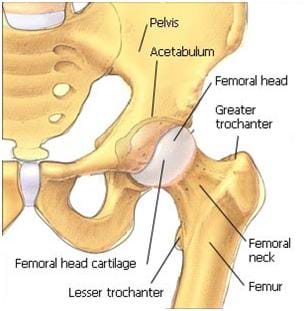
3d illustration of hip bone diagram hip bone anatomy. The bone surfaces of the femoral head and acetabulum have a smooth durable layer of articular cartilage that cushions the ends of the bones and allows for smooth movement. By natalia kremenon january 21, 2021in wiring diagram231 views. The foot bones shown in this diagram are the talus, navicular, cuneiform, cuboid, metatarsals and calcaneus. High resolution textures and displacement included.
The Artificial Hip Joint Valuable Information Before The Op Artiqo Endoprothetik from artiqo.de When the leg is stretched out, the knee joint is placed on a straight line with the hip and ankle (left). 3d illustration of hip bone diagram hip bone anatomy. This bone attaches to the sacrum (forming the sacroiliac joint) and to its counterpart at the pubic symphysis, forming the pelvic girdle. Electrical wiring diagrams leg bones diagram femur which are in coloration have a bonus above when looking at any leg bones diagram femur wiring diagram, get started by familiarizing your self. Hip anatomy pictures function problems treatment. Written by jupiterz saturday, march 25, 2017 add comment edit. Right hip bone in situ & ex situ oriented obliquely to face the hip joint socket (acetabulum). I'm not sure of what you mean by bone diagram.
Your leg bones are the longest and strongest bones in your body.
The head of your femur fits into your hip socket and the bottom end connects to your knee. Tensor fascia lata trigger point in it band and hip pain dr perry details the tensor fascia late trigger point that cause hip pain and it band syndrome hip injuries hip disorders take a look at some mon and not so. 2006 kia optima belt diagram. The knee joint is the largest joint in the body and is primarily a hinge joint, although some sliding and rotation occur. The foot bones shown in this diagram are the talus, navicular, cuneiform, cuboid, metatarsals and calcaneus. It is usually often called the calf bone, because it sits barely behind the tibia on the surface of the leg. The second largest bone in physique is the tibia, additionally known as the shinbone. Start studying leg bone diagram. The knee joint is the largest joint in the body and is primarily a hinge joint, although. The hip bone (os coxae, innominate bone, pelvic bone or coxal bone) is a large irregular bone, constricted in the center and expanded above and below. Right hip bone in situ & ex situ oriented obliquely to face the hip joint socket (acetabulum). At the distal end of the femur, two rounded condyles meet the tibia and fibula bones of the lower leg to form the knee joint. Human left hand bone parts names.
The hip joint is a ball and socket synovial type joint between the head of the femur and acetabulum of the pelvis leg bone diagram. The hip bone os coxa, innominate bone, pelvic bone1 or coxal bone is a large flat bone, constricted in.


0 Komentar